
INCIC cases ·
Four chambers at 83: what is your diagnosis?
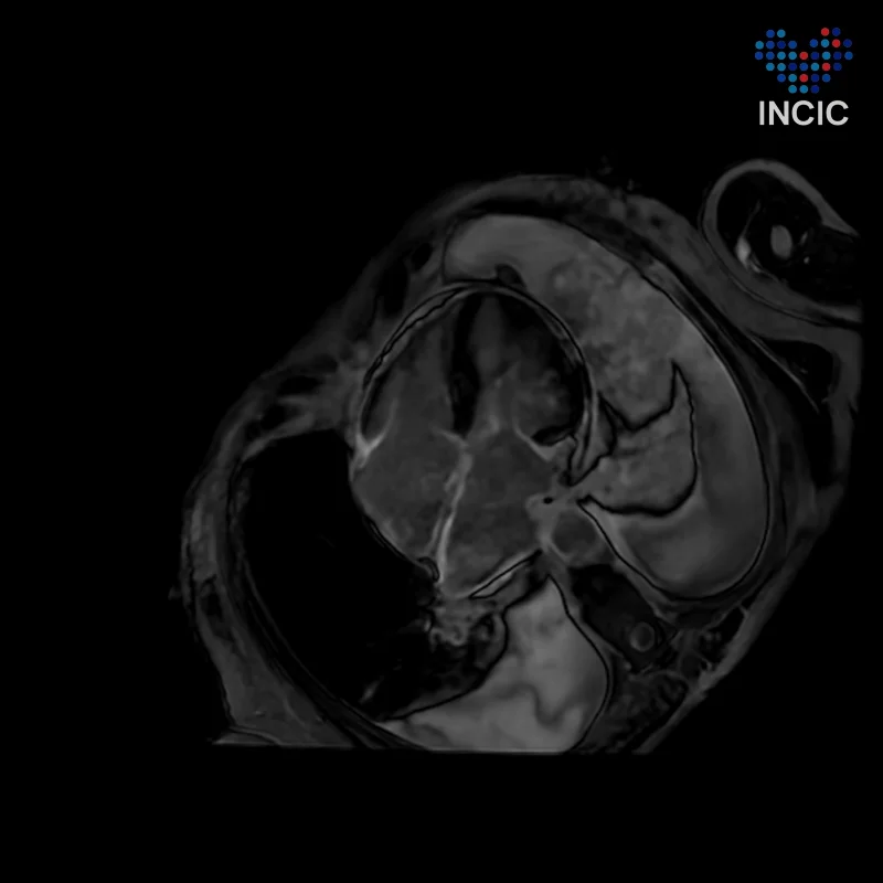
An 83-year-old woman investigated for dyspnoea. Cardiac MR, four-chamber cine.
Case by Julián Vega Adauy · jvega@incic.org
AtriaMild LV hypertrophyPleural effusion
What is your diagnosis?
Answer
Cardiac amyloidosis with isolated bi-atrial involvement
AmyloidosisIsolated atrial amyloidosis
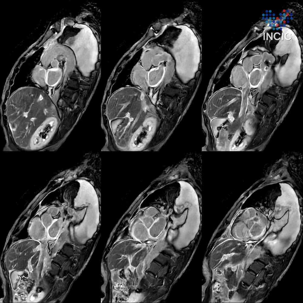
Both atria are dilated, with thickened walls and almost no contraction: the left atrial ejection fraction is 10%. Late enhancement is extensive and intense throughout both atria and the interatrial septum, while the left ventricle, with mild hypertrophy (septum 12.5 mm) and an LVEF of 65%, shows no focal fibrosis or oedema. The answer video shows filling with 4D Flow.
Pearls
- Look at the atria, not just the ventricle. Here the disease sits almost entirely in the atria: thickened, intensely enhancing and without function. The left ventricle shows only mild hypertrophy.
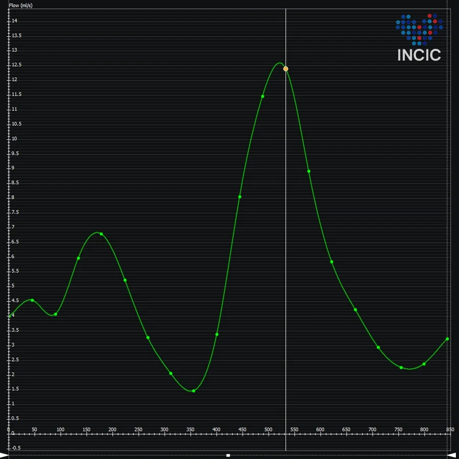
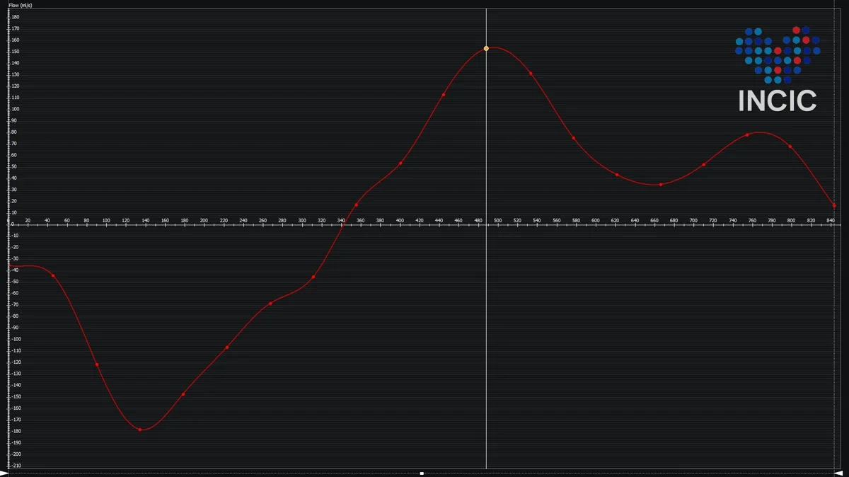
- Flow tells the physiology. With 4D Flow, pulmonary vein filling shows an S wave much smaller than the D wave, and mitral inflow shows a restrictive pattern. These are high filling pressures in an atrium that no longer contracts.
- The valves follow the annuli. Annular dilatation causes mild to moderate mitral regurgitation (RF 21%) and moderate tricuspid regurgitation (RF 37%), both functional.
- An ECV in the grey zone. Native T1 of 1067 ms and ECV of 33%, mildly raised. There is no categorical myocardial involvement, but early involvement cannot be ruled out at these values.
- Which amyloid. A pyrophosphate SPECT can help distinguish transthyretin (wild-type) amyloidosis from atrial natriuretic factor (ANP) deposition.
- What it rules out. The study rules out other causes of ventricular hypertrophy, such as sarcomeric hypertrophic cardiomyopathy and storage diseases.