
INCIC ·
4D flow: Dr. Javier López-Opitz shows how cardiac MR lets us see blood in motion
At the University Diploma in Cardiovascular Imaging by Bioimágenes, the INCIC specialist presented a case showing the clinical value of 4D flow for studying congenital heart disease without invasive procedures.
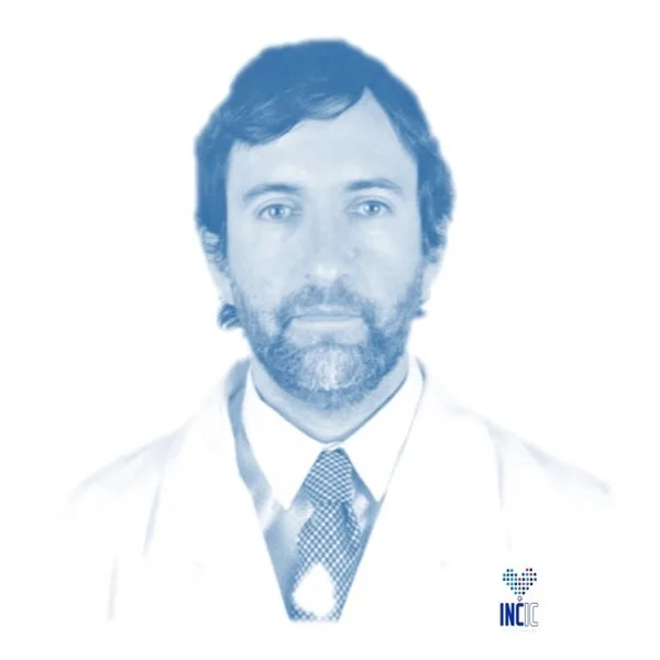
A routine echocardiography in a 25-year-old revealed a patent ductus arteriosus: a connection between the aorta and the pulmonary artery that normally closes in the first days of life. With that case, Dr. Javier López-Opitz, a cardiologist at INCIC, showed on Friday October 9, 2026, at the University Diploma in Cardiovascular Imaging by Bioimágenes, the clinical value and real-world impact of 4D flow, a cardiac magnetic resonance technique that enables advanced diagnosis of congenital heart disease without invasive procedures.
Seeing blood in motion
After the echocardiography, a cardiac MR was performed to answer three questions: how much blood flows through that connection, how the size and function of the heart look, and whether there are other associated abnormalities.
4D flow records how blood moves inside the heart and great vessels with every heartbeat, in three dimensions. This makes it possible to measure how much blood is diverted through the ductus without the need for cardiac catheterization.
Dr. López-Opitz also presented a comparison of the same analysis performed with two different processing programs.