How the heart sits in the chest, what the cardiac axes are, and how the LV walls and 17 segments are named.
Topography
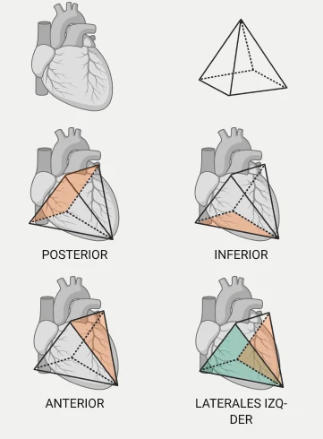
An inverted pyramid
•The heart has the shape of an inverted pyramid within the chest. Each face of the pyramid relates to an anatomical structure.
•Posterior: the base, facing the pulmonary veins and the oesophagus.
•Inferior: resting on the diaphragm.
•Anterior: in contact with the sternum.
•Lateral: facing the lungs (left and right).
Quick question
Which face of the heart rests on the diaphragm?
Cardiac segmentation
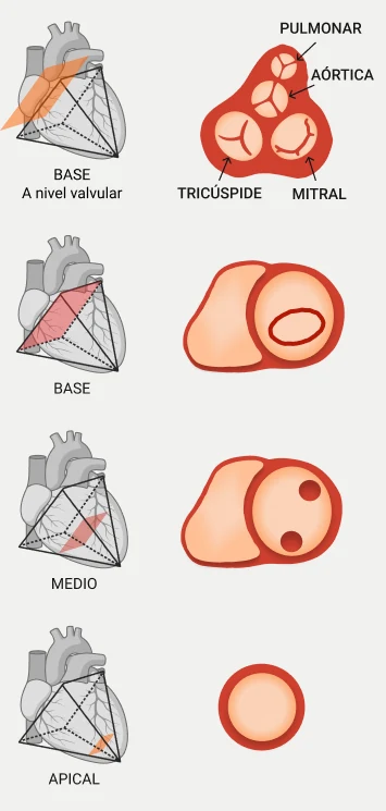
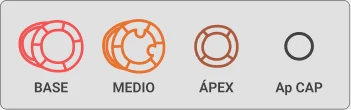
Three levels: basal, mid and apical
•From base to tip, the left ventricle is divided into three levels: basal, mid and apical.
•The base is at valve level, where the pulmonary, aortic, tricuspid and mitral valves sit.
•The papillary muscles insert at the mid level. The apical level is the tip of the heart.
Quick question
At which level are the cardiac valves?
Cardiac segmentation
What are the cardiac axes?
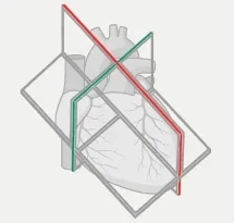
•Cardiac imaging uses planes that are oblique to the strict anatomical axes of the body (axial, coronal and sagittal).
•This is because the heart has its own orientation within the chest, and the views must align with its true axis.
•These planes are the basis of CMR planning and let us apply the AHA 17-segment model from different angles.
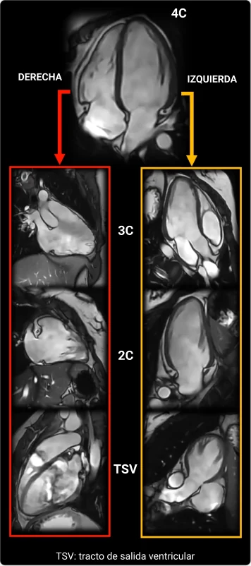
•Long axis: runs through the heart from base to apex, aligned with the long axis of the LV and following the interventricular septum. It includes the 2C, 3C and 4C.
•Short axis: perpendicular to the long axis; it slices the ventricle from base to apex and meets the septum at 90°.
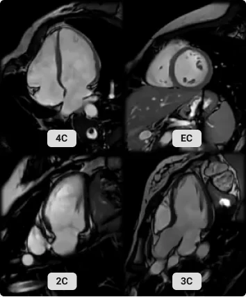
How they look on CMR: 4C, short axis (EC in Spanish), 2C and 3C.
Quick question
Why does cardiac MR use oblique planes instead of strict axial, coronal and sagittal?
Quick question
How does the short axis relate to the long axis?
Long axes
Left and right long axes
•Like the left side, the right heart has its own long axes: 3C, 2C and outflow tract.
•The 4C is the only long axis shared by the left and right sides.
Which long axis is shared by the left and right sides?
Cardiac segmentation
The three myocardial layers
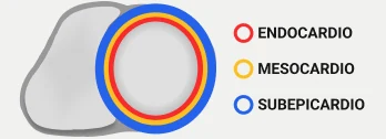
•Heart muscle is organised in three concentric layers:
•Endocardium: the innermost, in contact with the ventricular cavity.
•Mesocardium: the middle layer, where contractile fibres concentrate.
•Subepicardium: the outer layer, next to the pericardium.
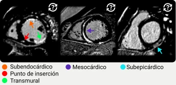
•On CMR, the layer involved helps tell diseases apart: the subendocardium is typically affected in infarction (ischaemic pattern), the subepicardium is characteristic of myocarditis and the mesocardium is common in non-ischaemic cardiomyopathy.
Late gadolinium enhancement by layer involved.
Quick question
Subendocardial late enhancement typically suggests…
LV walls
Four walls and six segments
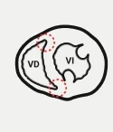
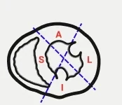
•The right ventricle (RV) attaches to the left ventricle (LV) at the superior and inferior interventricular insertion points.
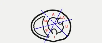
•Lines drawn from each insertion point through the middle of the LV cavity (in diastole) divide the LV into 4 walls: anterior (A), septal (S), inferior (I) and lateral (L). The septum is so called because it separates both ventricles.
•Only the septal and lateral walls, being larger, are split again in two, each half named after the nearest wall: anteroseptal and inferoseptal, anterolateral and inferolateral.
📝 The figures use Spanish abbreviations, which name the divided wall first: SA septal-anterior (anteroseptal), SI septal-inferior (inferoseptal), LA lateral-anterior (anterolateral), LI lateral-inferior (inferolateral).
Quick question
Which LV walls are split into two segments?
AHA model
The 17 segments
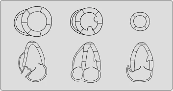
•The AHA (American Heart Association) standard divides the LV myocardium into 17 segments, grouped into three levels from base to apex:
•Basal (1–6): closest to the AV valves. It ends just before the papillary muscles.
•Mid (7–12): the middle portion, where the papillary muscles insert.
•Apical (13–16): the distal end, the tip. The cavity narrows here and the septal and lateral walls are not split.
•True apex (17): the most distal point of the LV, a virtual slice that does not touch the cavity.
📝 Why a "virtual" 17th segment? The original model had 16 segments; segment 17, the apical cap, was added to align segmentation with nuclear medicine.
Quick question
At which level do the papillary muscles insert?
Quick question
How many segments does the apical level have?
AHA model
Segments from different axes
•The 17 segments can be seen from several imaging planes:
•Short axis (SAX): shows the segments in slices across the long axis of the heart.
•Long axes (2C, 3C, 4C): show longitudinal projections. Each view cuts a different combination of segments.
•This multiplanar correlation is key to interpreting cine, perfusion, parametric mapping and late enhancement findings correctly.
💡 In the next lesson you will see which segments each long axis cuts.
Quick question
Which plane shows the segments in slices across the heart’s axis?
🏁
Lesson complete
0
XP
0
Right on first try
📌 Remember
•Heart as an inverted pyramid: inferior face on the diaphragm
•Oblique cardiac axes: long axis (2C, 3C, 4C) and perpendicular short axis
•4C, the only long axis shared by left and right sides
Content based on CMR Essential (J. Vega) and the SCMR standardised protocols (Kramer et al., JCMR 2020).
Visit and ad measurement
With your permission we use Google Analytics and Google Ads to count visits to the site and find out whether our ads bring enquiries. We do not personalise ads or send your form data to Google. Learn more